facs flow cytometry protocol
Anti-Neu5Gc Antibody Kit Protocol. The flow cytometry protocols below provide detailed procedures for the treatment and staining of cells prior to using a flow cytometer.
How Does Flow Cytometry Work Nanocellect
Flow Cytometry FCM and FACS protocols.

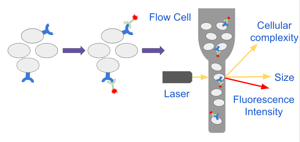
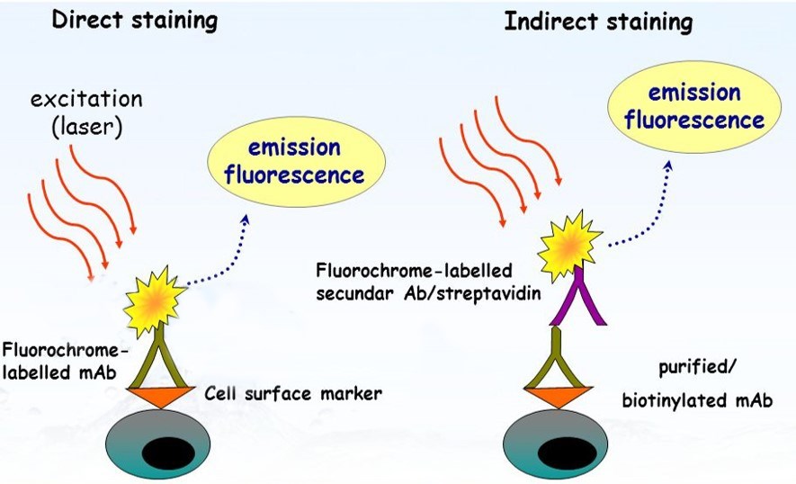
. Add 01-10 μgml of the primary labeled antibody. Direct staining of cells applicable where the fluorophore is. The following flow cytometry.
Request a quote and see how Agilent has advanced the boundaries of flow cytometry. Flow cytometry is the measurement of chemical and physical properties of cells as they flow one by one through an integration point most commonly a laser. Ad Compatible with optional autosamplers and robotic integration for walk-away automation.
Ad MALS verified high binding ability. Use this buffer also for all washes until directed to use Sorting Buffer Adjust. Run sticky samples at high flow rates with a system that is exceptionally clog resistant.
Incubate on ice for 5 minutes. Precision Count Beads Protocol and Applications. Flow Cytometry FACS Protocols PSR The BD FACSCalibur platform allows users to perform both cell analysis and cell sorting in a single benchtop system.
MALS verified heterodimers with various tags. EdU 5-ethynyl-2-deoxyuridine is a nucleoside analog to thymidine and is incorporated into DNA. Ad Compatible with optional autosamplers and robotic integration for walk-away automation.
EV Precipitation Solution 4 um beads for. This protocol has two major steps. Please refer to the APPLICATIONS section on the front page of product datasheet or product webpage to determine if this product is validated and approved for use in Flow.
Centrifuge for 5 minutes at 350xg and discard supernatant. Streamline EV Flow Cytometry with EV-FACS. Perform red blood cell lysis per lab protocol either ACT ACK or LSM.
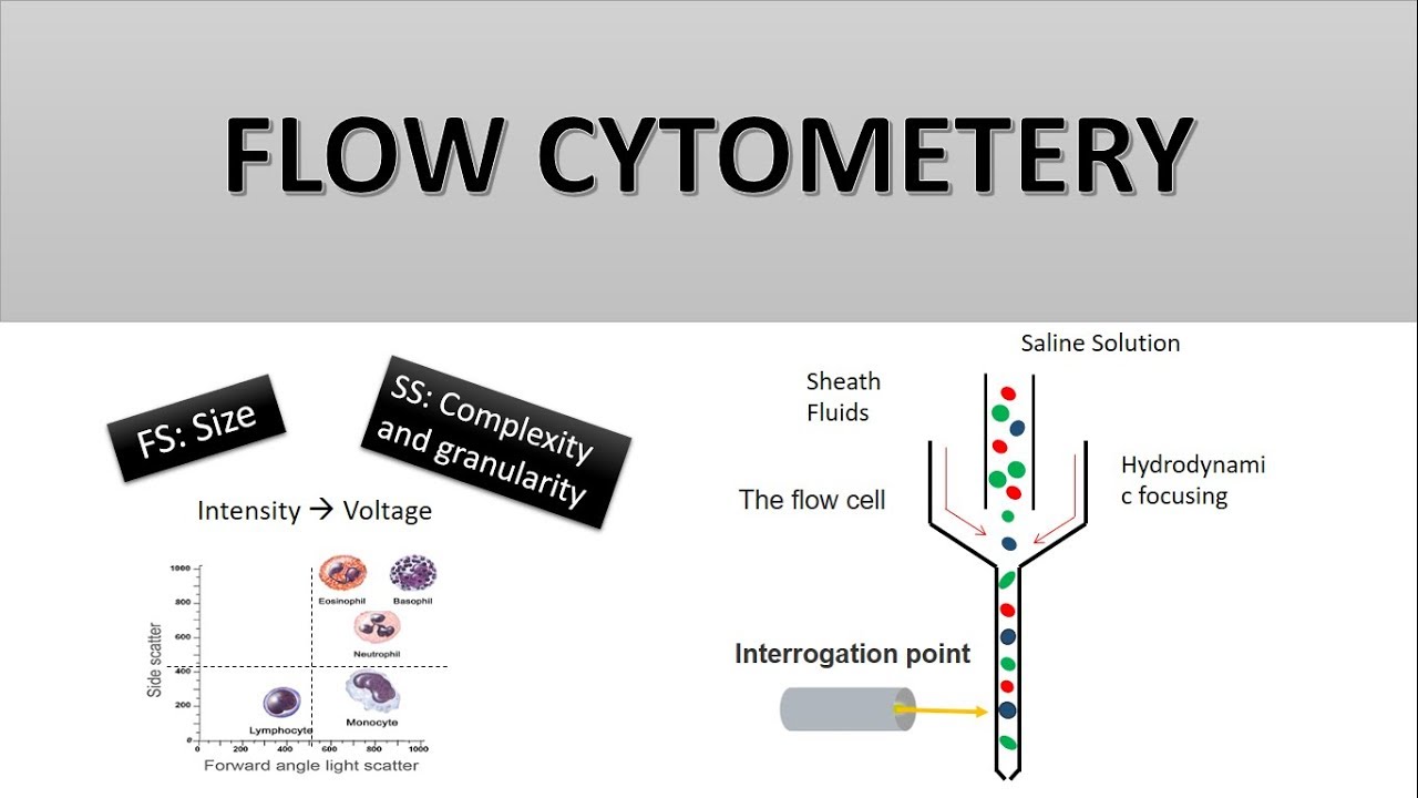
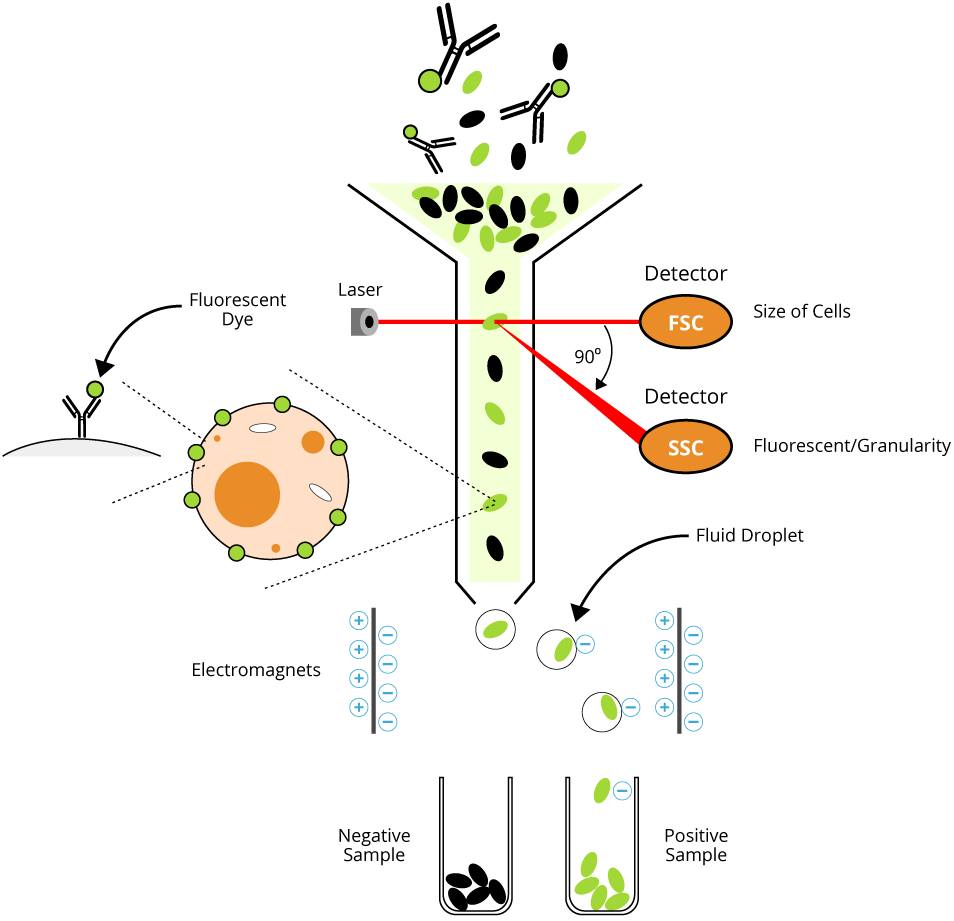
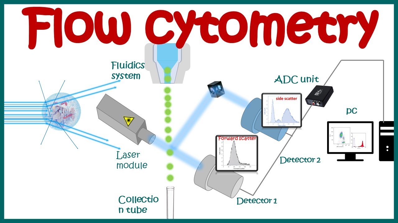
Flow cytometry FACS staining protocol Cell surface staining 1. The system supports a wide. Flow cytometry FCM is a means of measuring certain physical and chemical characteristics of cells or particles as they pass in a.
As cells scatter laser light in. Repeat wash as in step 2. The Intacellular Flow Cytometry Staining Protocol describes the process for intracellular staining of various cell types in vivo-stimulated tissues in vitro-stimulated cultures and whole blood.
This protocol describes an efficient methodology for generating knock-out and knock-in clonal populations of human induced pluripotent stem cells hiPSCs using CRISPR-Cas9 technology. Cell Surface Flow Cytometry Staining Protocol. MALS verified heterodimers with various tags.
The Click-iT EdU Flow Cytometry Assay Kits are novel alternatives to the BrdU assay. Harvest wash the cells single cell suspension and adjust cell number to a concentration of 1-5106 cellsml in ice cold FACS. Run sticky samples at high flow rates with a system that is exceptionally clog resistant.
Ad Learn how Agilent NovoCyte Flow cytometers have advanced the boundaries of flow cytometry. Get information on stimulation of cells appropriate cultures for generating human mouse and rat. Re-suspend in FACS staining buffer.
First we prepare a. This protocol was optimized for use with mouse mammary tumors where immune cells may represent 20-40 of total cells. Stop cell lysis by adding 10ml Cell Staining Buffer to the tube.
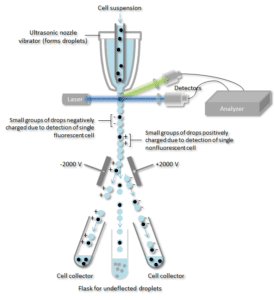
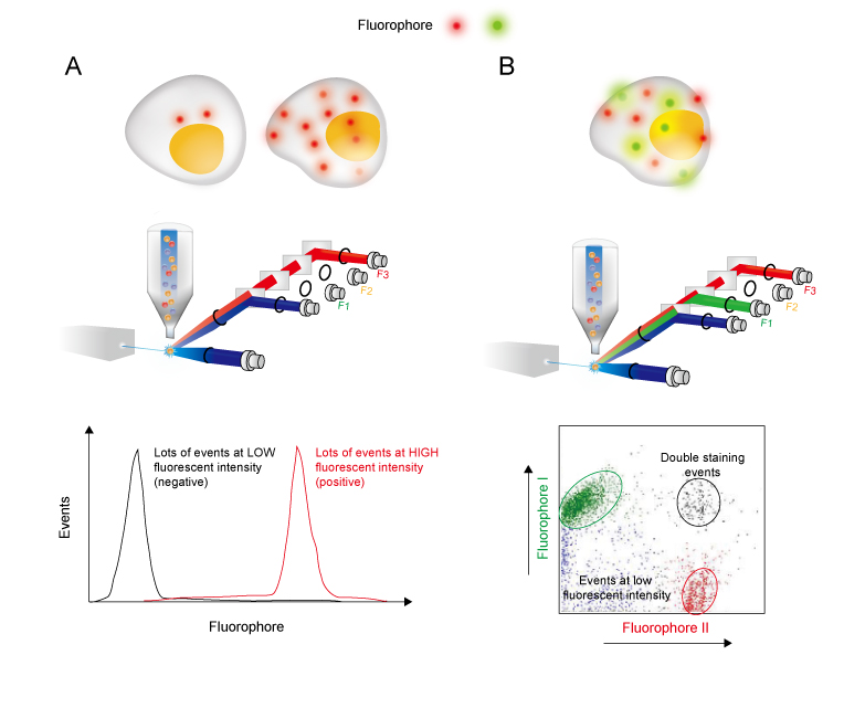
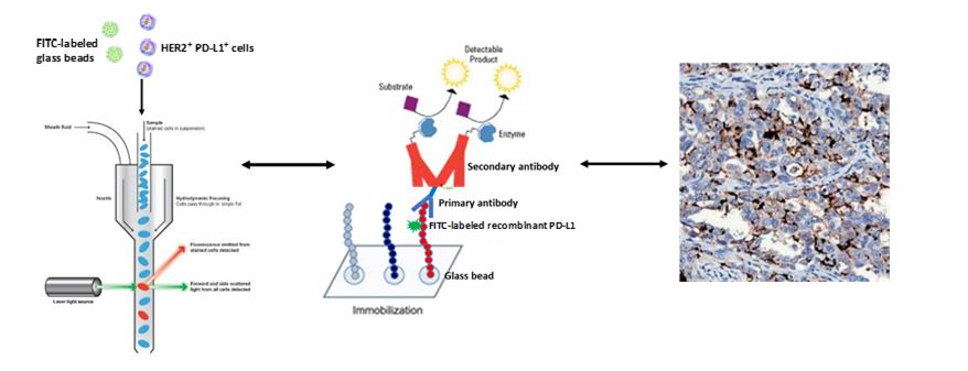
Incubate for at least 30 min at room temperature or 4C in the dark. By utilizing highly specific antibodies labeled with fluorescent conjugates FACS analysis allows us to simultaneously collect data on and sort a biological sample by a nearly limitless number. Immunofluorescent Staining of Intracellular Cytokines for Flow Cytometric Analysis.
Ad MALS verified high binding ability. Dilutions if necessary should be made in FACS buffer. Everything needed to isolate and analyze extracellular vesicles by flow cytometry including.
Flow Cytometry is used for research applications such as immunophenotyping DNA studies cell cycle analysis and fluorescence-activated cell sorting FACS.
In The Protocol Developed By Bernhard Fuchs S Team Bacterial Groups Are Enriched In Three Steps 1 In Situ Hybridization Postdoctoral Researcher Microbiology

Flow Cytometry Detection Of Surface And Intracellular Antigens In Pancreas From A Single Mouse Embryo Star Protocols

Single Cell Rna Expression Analysis Using Flow Cytometry Based On Specific Probe Ligation And Rolling Circle Amplification Acs Sensors
Flow Cytometry And Cell Sorting By Facs In The Flow Cell 1 The Download Scientific Diagram
Flow Cytometry Guide Creative Diagnostics
The Principle Of Flow Cytometry And Facs 2 Facs Fluorescence Activated Cell Sorting Youtube
Analyzing Single Cells With Flow Cytometry
Flow Cytometry Protocols

Flow Cytometry Creative Biolabs

Flow Cytometry Based Protocols For Human Blood Marrow Immunophenotyping With Minimal Sample Perturbation Star Protocols

Optimized Flow Cytometric Protocol For The Detection Of Functional Subsets Of Low Frequency Antigen Specific Cd4 And Cd8 T Cells Sciencedirect
Flow Cytometry Basic Principles What The Use Of Flow Cytometry Cell Sorting By Facs Youtube

Diagnostic Potential Of Imaging Flow Cytometry Trends In Biotechnology
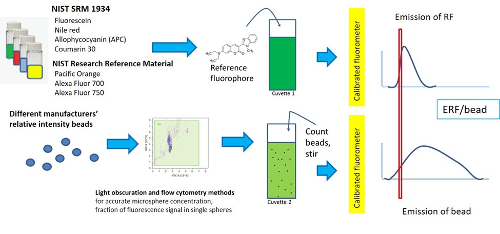
Quantitative Flow Cytometry Measurements Nist

Flow Cytometry Sample Preparation Proteintech Group

Flow Cytometry Facs Protocols Sino Biological
Quantitative Flow Cytometry Measurements Nist

Purification Of Micronuclei From Cultured Cells By Flow Cytometry Star Protocols
Direct Staining Flow Cytometry Creative Biolabs